Reproductive System Female Dog: Understanding Health and Care Tips

Reproductive system female dog. Understanding the reproductive system of female dogs reveals a complex biological framework that not only enables procreation but also reflects evolutionary adaptations pivotal for species survival. Female dogs, scientifically referred to as bitches, exhibit a remarkable reproductive system designed to accommodate multiple offspring in a single pregnancy, highlighting their status as litter-bearing creatures.
Anatomy and Functionality: Key Components of the Canine Reproductive System

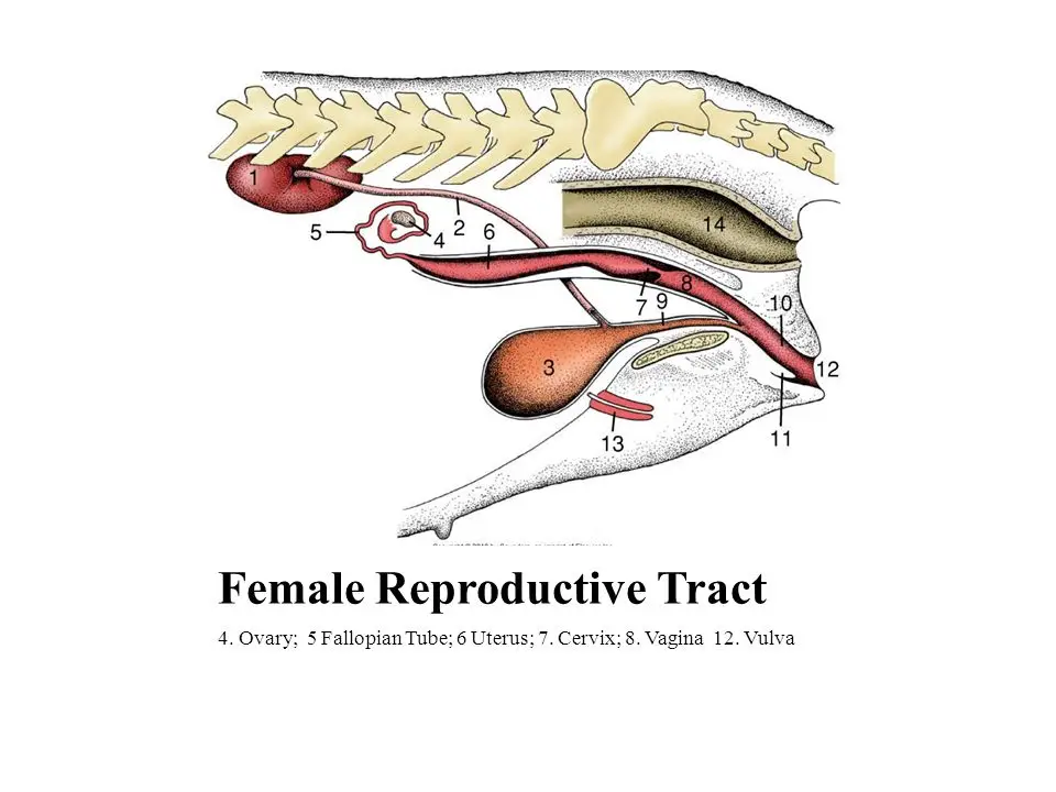
A closer look at the anatomy reveals essential components: ovaries, oviducts, uterus, cervix, and vagina. The ovaries are responsible for producing ova and hormones that regulate the estrous cycle and support pregnancy. The oviducts serve as the passageway for ova post-ovulation, where fertilization can occur if sperm are present. The uterus, notable for its capacity to expand and host developing embryos, is structured to accommodate multiple fetuses, reflecting the evolutionary advantage of birthing litters rather than single offspring. This characteristic amplifies survival odds for the young. An equally fascinating aspect is the interaction of hormonal pathways governing these processes. Follicle-stimulating hormone (FSH) spurs follicle development in the ovaries, while luteinizing hormone (LH) prompts ovulation.
Reproductive System Female Dog – Ovaries: The Powerhouses of Reproduction

The ovaries are the primary reproductive organs in female dogs, responsible for the production of ova (eggs) and the secretion of essential hormones. These small, almond-shaped structures are located in the abdominal cavity, near the junction of the uterus and the oviducts.
The ovaries undergo cyclical changes throughout the estrous cycle, which is marked by significant hormonal fluctuations. During the proestrus and estrus phases, the ovaries respond to the surge of follicle-stimulating hormone (FSH) by developing and maturing follicles, each containing a single ovum. As the cycle progresses, the luteinizing hormone (LH) triggers the release of the mature ova from the ovarian follicles, a process known as ovulation.
The ovaries also produce the hormones progesterone and estrogen, which play crucial roles in regulating the estrous cycle, preparing the uterus for implantation, and supporting the maintenance of pregnancy. These hormonal variations not only influence the dog’s reproductive capacity but also impact her overall behavior and physiology.
Oviducts: Highways for Fertilization

The oviducts, also known as the fallopian tubes, are the conduits through which the ova (eggs) travel from the ovaries to the uterus. These slender, muscular structures are approximately 10-15 cm long and are lined with ciliated epithelial cells that facilitate the movement of the ova.
After ovulation, the ova are captured by the fimbriated end of the oviduct and begin their journey towards the uterus. If sperm are present in the oviduct, fertilization can occur, and the zygote (fertilized egg) will continue its descent into the uterus, where it will implant and develop into an embryo and fetus.
The oviducts play a crucial role in the reproductive process, as they provide a safe passage for the ova and ensure that fertilization can take place in the appropriate environment. Any disruption or abnormality in the structure or function of the oviducts can significantly impact the female dog’s fertility and reproductive success.
Uterus: The Nurturing Womb

The uterus, or the “womb,” is the central reproductive organ in the female dog, responsible for housing and nourishing the developing offspring during pregnancy. This muscular, pear-shaped structure is located in the pelvic cavity and is composed of two distinct parts: the body of the uterus and the two uterine horns.
The uterine horns are particularly remarkable in female dogs, as they are adapted to accommodate multiple fetuses during pregnancy. This characteristic is a reflection of the species’ evolutionary strategy, as litter-bearing breeds tend to have a higher reproductive capacity and increased chances of survival for their offspring.
The uterine wall consists of three layers: the endometrium, the myometrium, and the perimetrium. The endometrium is the innermost layer, which undergoes cyclical changes to prepare for the implantation of the fertilized egg. The myometrium, the middle layer, is composed of smooth muscle fibers that contract during parturition (birth) to expel the puppies. The outermost layer, the perimetrium, is a serous membrane that helps to protect and support the uterus.
The uterus plays a vital role in the reproductive process, providing a nurturing environment for the developing fetuses and facilitating the delivery of the puppies during parturition. Any abnormalities or disorders affecting the uterus, such as pyometra (a potentially life-threatening uterine infection), can have significant consequences on the female dog’s reproductive health and overall well-being.
Cervix and Vagina: Gateways to Conception and Birth

The cervix and vagina are the final components of the female dog’s reproductive system, serving as the passageways for sperm entry during mating and the exit route for the puppies during parturition.
The cervix is a narrow, muscular structure that connects the uterus to the vagina. During the estrous cycle, the cervix undergoes changes in its consistency and position, allowing it to open and close in response to hormonal fluctuations. This function is crucial, as it regulates the entry of sperm during mating and the passage of the puppies during birth.
The vagina, on the other hand, is a tubular structure that extends from the cervix to the external genitalia. It serves as the site of semen deposition during mating and the birth canal through which the puppies are delivered. The vagina is lined with a mucous membrane that lubricates and protects the delicate tissues during intercourse and parturition.
The coordination between the cervix and the vagina is essential for successful reproduction. Any abnormalities or infections in these structures can lead to complications, such as difficulty in breeding or obstructed labor, which can compromise the health and well-being of both the female dog and her offspring.
Reproductive Cycle: Orchestrating Fertility and Gestation

The reproductive cycle of the female dog, known as the estrous cycle, is a complex and precisely timed series of events that prepares the dog’s body for breeding, pregnancy, and the subsequent nurturing of the offspring. This cycle is marked by significant hormonal changes that impact the dog’s behavior, physiology, and reproductive capacity.
The estrous cycle in dogs is typically divided into four distinct phases: proestrus, estrus, diestrus, and anestrus. Each phase is characterized by unique hormonal profiles and corresponding physiological and behavioral changes.
Proestrus: The Prelude to Fertility

The proestrus phase is the initial stage of the estrous cycle, typically lasting 7-10 days. During this phase, the levels of estrogen begin to rise, leading to the development and maturation of the ovarian follicles. This hormonal change triggers the swelling of the vulva and the onset of vaginal bleeding, which is often the first visible sign of the dog’s heat cycle.
During proestrus, the female dog may exhibit increased affection, restlessness, and even aggression towards other dogs, as her body prepares for the impending ovulation and potential mating. It is important to note that during this phase, the female dog is not yet receptive to mating, and any attempted breeding should be discouraged to prevent unwanted pregnancies.
Estrus: The Fertile Phase

The estrus phase, also known as the “heat” or “standing heat,” follows proestrus and typically lasts 5-13 days. This is the stage during which the female dog becomes receptive to mating and is at the peak of her fertility.
During estrus, the levels of estrogen continue to rise, and the female dog exhibits clear behavioral changes. She may become more affectionate, seek out male dogs, and assume a characteristic “lordosis” posture to signal her readiness for breeding. The vulva also becomes less swollen, and the vaginal discharge changes in color and consistency, becoming more clear and watery.
It is crucial to closely monitor the female dog during the estrus phase, as this is the optimal time for mating to occur. Breeding during this phase increases the chances of successful conception and pregnancy.
Diestrus: The Luteal Phase
The diestrus phase follows the estrus phase and typically lasts for 60-90 days. During this time, the levels of progesterone, the hormone responsible for maintaining pregnancy, begin to rise. If the female dog becomes pregnant, diestrus will continue throughout the gestation period, supporting the development and nourishment of the fetuses.
If the female dog does not become pregnant, the diestrus phase will still occur, but it will be shorter in duration and will eventually transition into the anestrus phase.
Anestrus: The Resting Phase
The anestrus phase is the final stage of the estrous cycle, and it is characterized by a period of reproductive quiescence. During this phase, the female dog’s reproductive system rests and prepares for the next cycle. Hormonal levels are low, and the dog exhibits no outward signs of heat or receptivity to mating.
The duration of the anestrus phase can vary, but it typically lasts for 2-4 months in most female dogs. This resting period allows the dog’s body to recover and rejuvenate before the next cycle begins.
Understanding the complexities of the estrous cycle is crucial for breeders and veterinarians in managing the reproductive health of female dogs. By monitoring the various stages and their corresponding hormonal and behavioral changes, they can better time breeding, ensure successful pregnancies, and provide appropriate care and support for the female dog and her offspring.
Implications and Broader Context: Connecting Health and Breeding Practices

The health of the reproductive system has vast implications—not only for successful breeding but also for the overall well-being of the female dog. Disorders affecting any component of the reproductive system, from pyometra (a potentially life-threatening infection of the uterus) to ovarian cysts, can hinder reproductive success and have dire consequences on the bitch’s health.
Regular veterinary check-ups and informed breeding practices become essential in sustaining both the breed standards and the individual pet’s quality of life. Breeders must be vigilant in monitoring the reproductive health of their breeding stock and addressing any underlying issues to ensure the production of healthy, viable offspring.
Moreover, as breeders aim for specific traits and lineage preservation, ethical considerations arise regarding overbreeding or inadequate health oversight, potentially resulting in inherited health issues that compromise long-term viability. This necessity for conscientious breeding evokes larger questions about animal husbandry ethics and the responsibilities held by those who care for these beloved companions.
In view of ongoing research surrounding canine reproduction, there are opportunities for further engaging with this subject—be it through improved artificial insemination techniques, enhanced understanding of embryonic development, or protective health protocols to better safeguard both the dam and her future puppies. Such advancements have the potential to enrich not just the lives of individual dogs but influence entire breeds, setting paradigms for humane and sustainable canine care.
Conclusion

The reproductive system of the female dog is a remarkable and intricate biological framework, shaped by evolutionary adaptations to ensure the survival and proliferation of the species. From the ovaries responsible for ova and hormone production to the uterus designed to accommodate multiple fetuses, each component of the canine reproductive system plays a vital role in the complex processes of breeding, gestation, and parturition.
By understanding the anatomy, functionality, and hormonal regulation of the female dog’s reproductive system, we can not only gain deeper insights into the biology of these beloved companions but also develop more effective and compassionate approaches to breeding and caregiving. As we continue to explore the nuances of canine reproduction, we unlock opportunities to enhance the health and well-being of dogs, fostering a future where responsible breeding practices and advancements in veterinary science converge to safeguard the flourishing of the canine species.


